|
Size: 1925
Comment:
|
Size: 1868
Comment:
|
| Deletions are marked like this. | Additions are marked like this. |
| Line 39: | Line 39: |
| Structural biology overview {{attachment:struct_bio.png||align="center",width=400}} |
Structural biology overview {{attachment:struct_bio.png||align="right",width=400}} |
| Line 42: | Line 41: |
| Transmission electron microscopes (TEM) {{attachment:tem.png||align="center",width=400}} |
Transmission electron microscopes (TEM) {{attachment:tem.png||align="right",width=400}} |
| Line 45: | Line 43: |
| TEM grids {{attachment:grid.png||align="center",width=400}} |
TEM grids {{attachment:grid.png||align="right",width=400}} |
| Line 48: | Line 45: |
| Movie-mode imaging {{attachment:movie.png||align="center",width=400}} |
Movie-mode imaging {{attachment:movie.png||align="right",width=400}} |
| Line 51: | Line 47: |
| Basic image processing {{attachment:imgproc.png||align="center",width=400}} |
Basic image processing {{attachment:imgproc.png||align="right",width=400}} |
| Line 54: | Line 49: |
| Fourier transforms {{attachment:fft.png||align="center",width=400}} |
Fourier transforms {{attachment:fft.png||align="right",width=400}} |
| Line 57: | Line 51: |
| Single particle analysis {{attachment:spa.png||align="center",width=400}} |
Single particle analysis {{attachment:spa.png||align="right",width=400}} |
| Line 60: | Line 53: |
| Contrast transfer function (CTF) {{attachment:ctf.png||align="center",width=400}} |
Contrast transfer function (CTF) {{attachment:ctf.png||align="right",width=400}} |
| Line 63: | Line 55: |
| Resolution measurement via Fourier Shell Correlation (FSC) {{attachment:fsc.png||align="center",width=400}} |
Resolution measurement via Fourier Shell Correlation (FSC) {{attachment:fsc.png||align="right",width=400}} |
| Line 66: | Line 57: |
| Electron cryo-tomography (cryoET) {{attachment:cryoet.png||align="center",width=400}} |
Electron cryo-tomography (cryoET) {{attachment:cryoet.png||align="right",width=400}} |
| Line 69: | Line 59: |
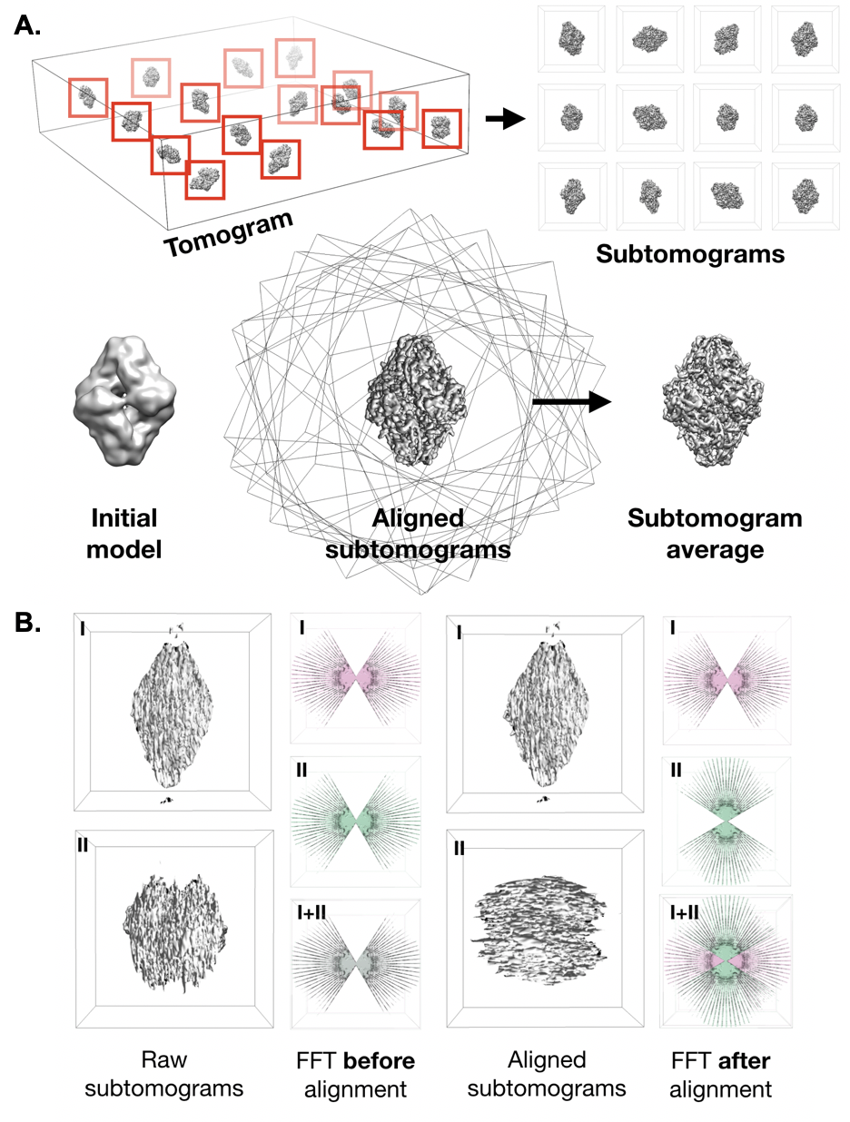
| Subtomogram Averaging / Single particle tomography (SPT) {{attachment:subtomoavg.png||align="center",width=400}} |
Subtomogram Averaging {{attachment:subtomoavg.png||align="right",width=400}} |
Michael Bell, Ludtke Lab (2014 - 2019)
All are welcome to use the materials provided here. Credit is appreciated but not required.
Thesis
Note: All protein illustrations in are licensed under a Creative Commons Attribution 4.0 International license by David S. Goodsell and the RCSB PDB.
Thesis Defense Presentation
Posters
cryoet_poster.pdf, cryoet_poster.pptx
Note, these posters are have been uploaded without modification. There may be typos.
Talks
microscopy_microanalysis_2018.ppt
Select Thesis Figures
Structural biology overview 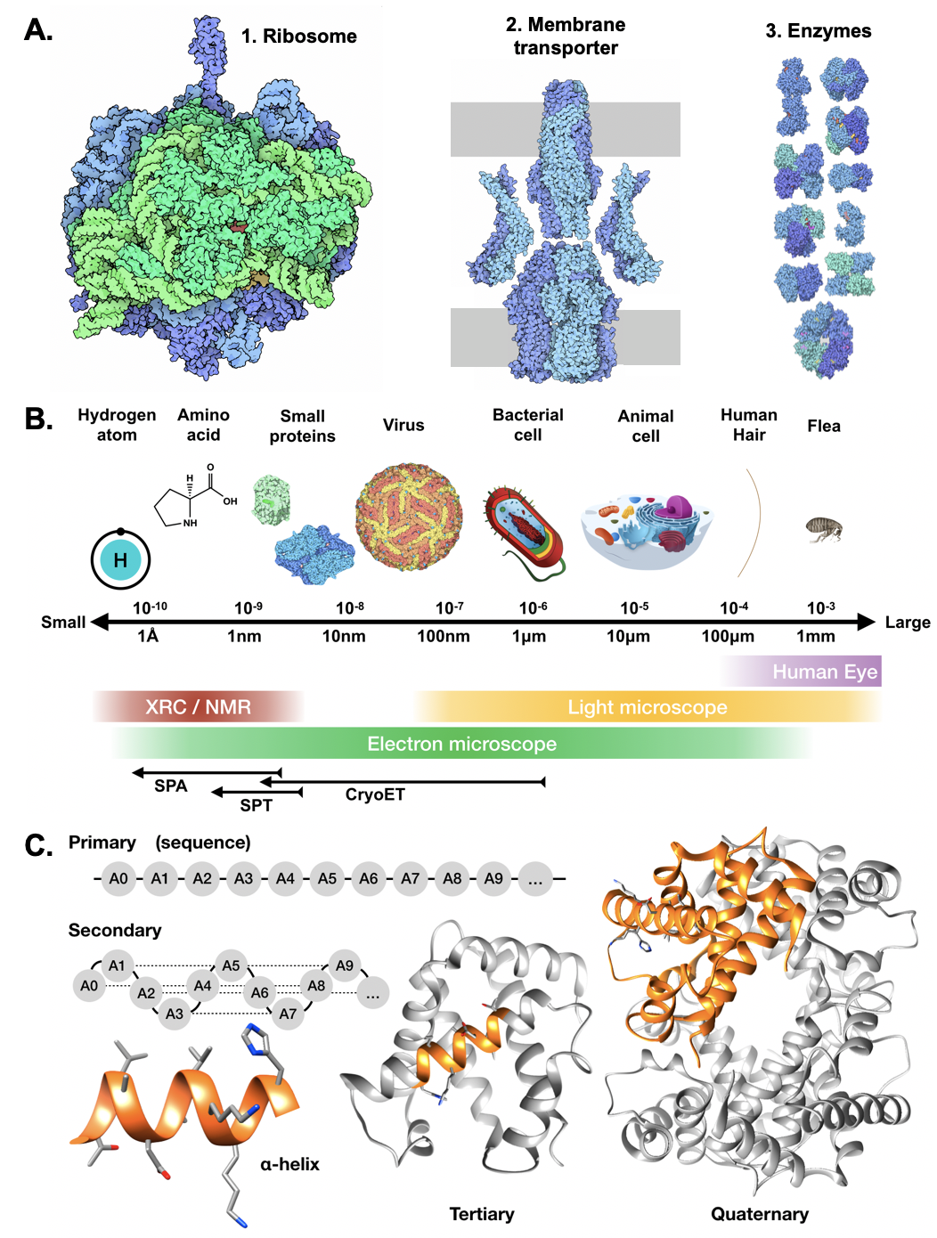
Transmission electron microscopes (TEM) 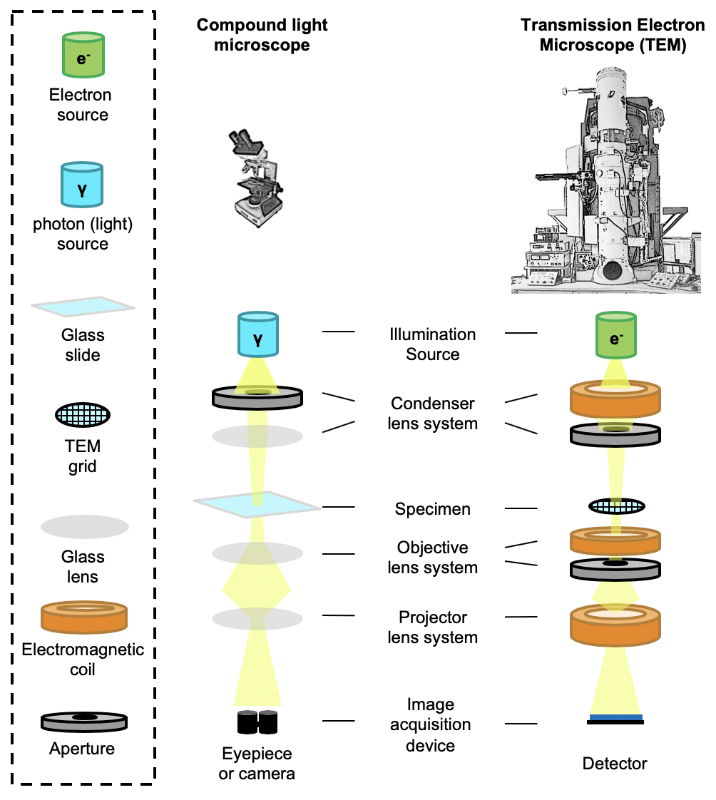
TEM grids 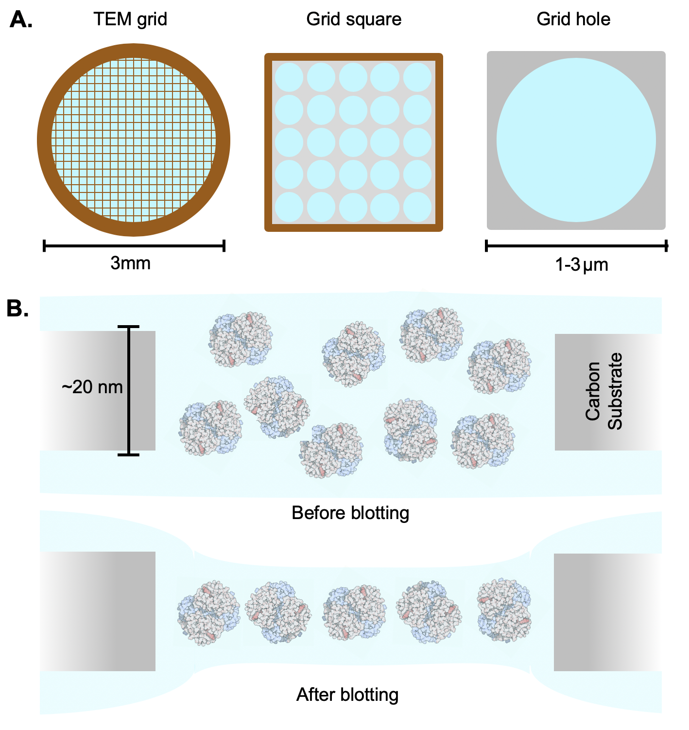
Movie-mode imaging 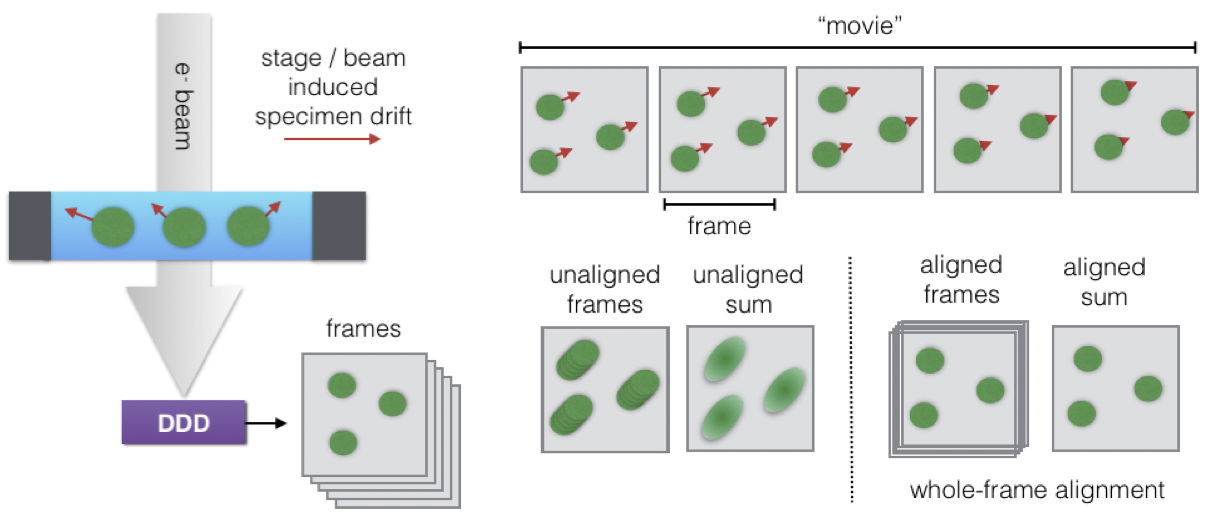
Basic image processing 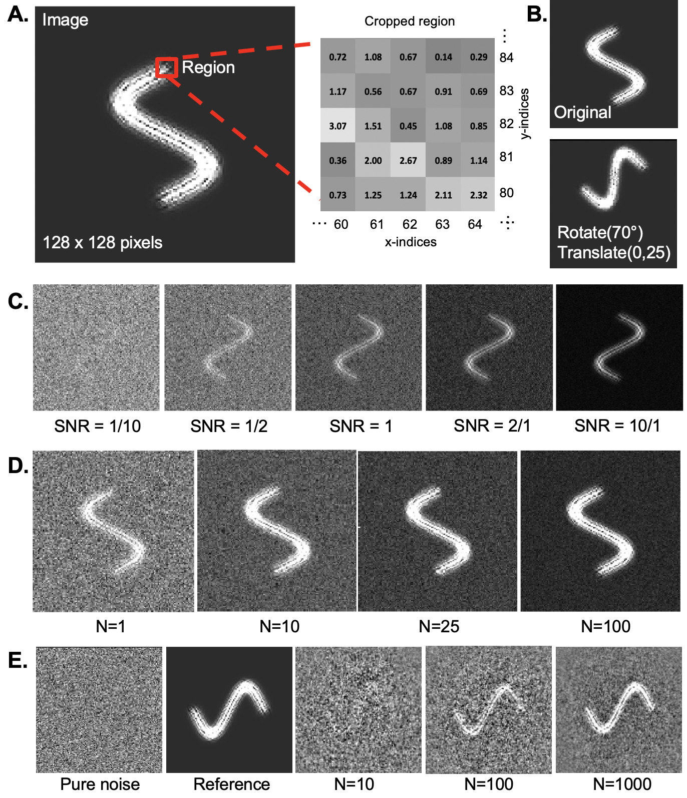
Fourier transforms 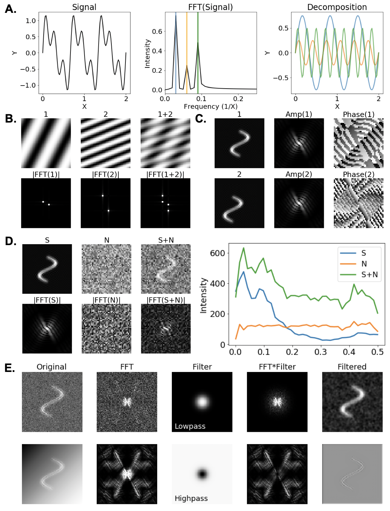
Single particle analysis 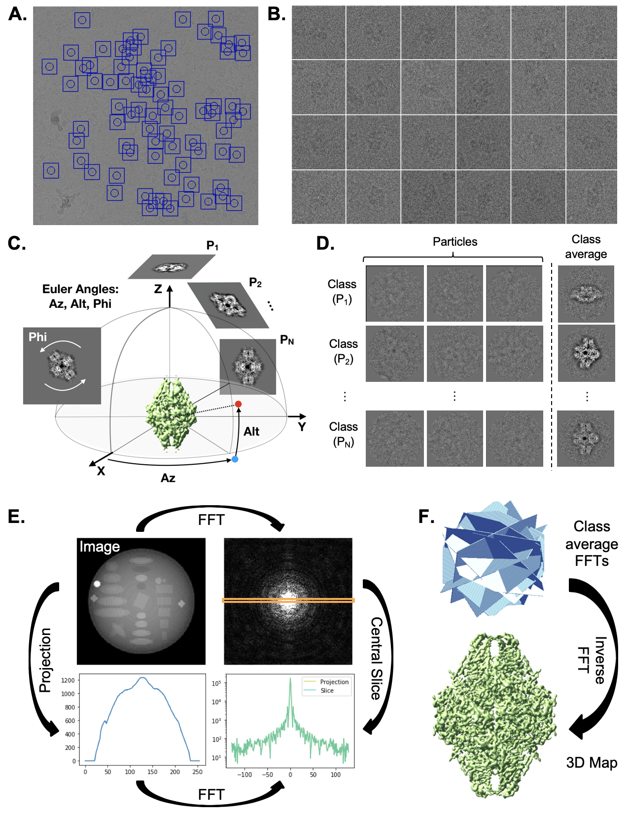
Contrast transfer function (CTF) 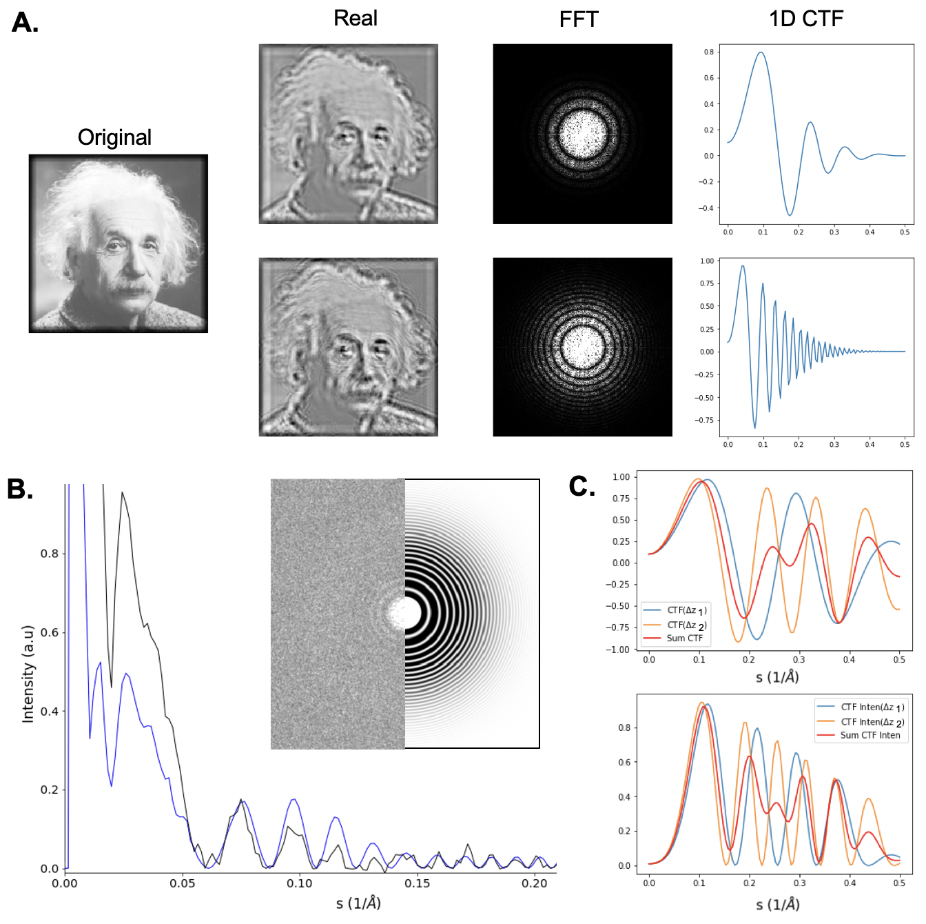
Resolution measurement via Fourier Shell Correlation (FSC) 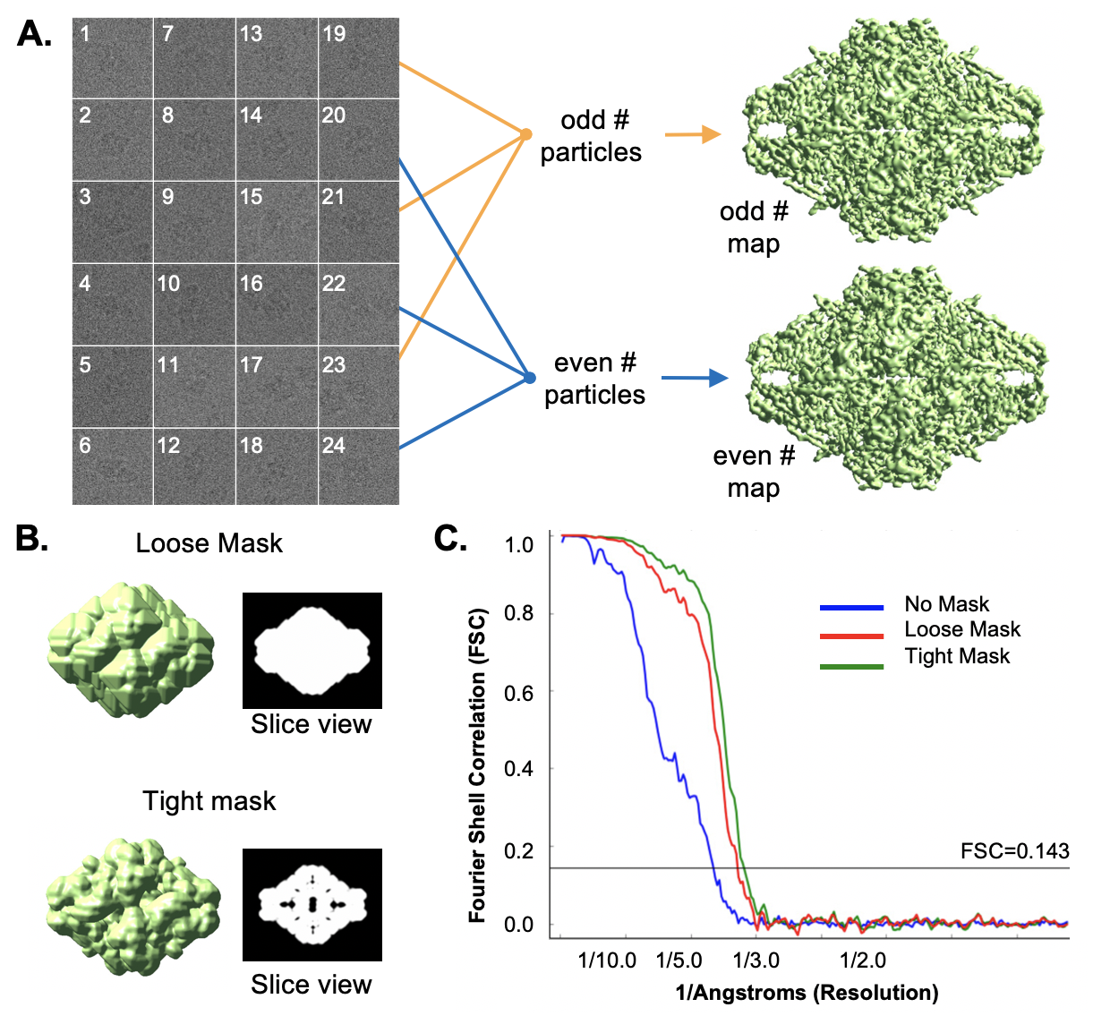
Electron cryo-tomography (cryoET) 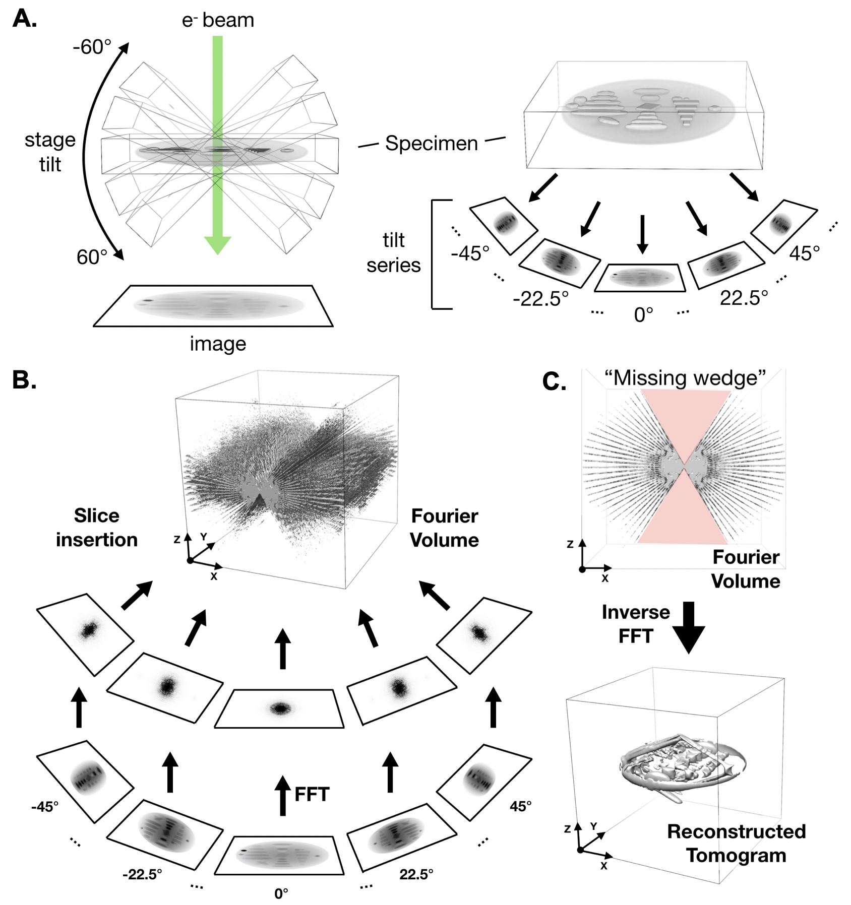
Subtomogram Averaging 